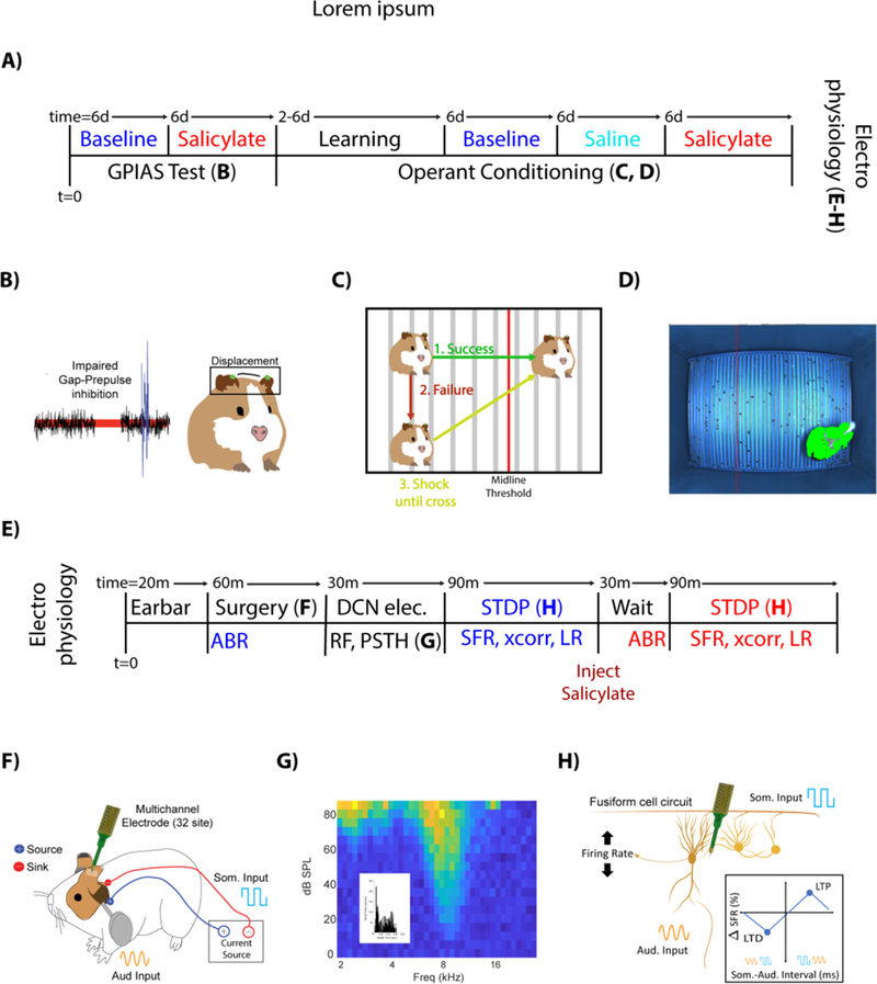
Figure 1. Experimental Design and Timeline.

A) Animals were tested for tinnitus baseline using the adapted GPIAS paradigm (see METHODS) 6 days (Mon/Thurs, or Tue/Fri) and with salicylate for 6 days. Following GPIAS, animals were trained to move when a sound was presented (2–6 days; Mon/Wed/Fri). Animals that successfully learned crossing received 6 days of baseline testing, followed by administration of saline and then salicylate, each for 6 days. After operant conditioning, DCN electrophysiology was performed. B) Tinnitus impairs gap-prepulse inhibition when spectrally like a background carrier band. Guinea pig pinna tips were painted green, tracked using high speed cameras and the pinna-startle displacement computed. C) If guinea pigs crossed the midline when a sound was introduced, no shock was given (Green). If they failed to cross the midline during a sound (Red), the guinea pig received a footshock until it crossed the midline (Yellow). D) Sample frame, with the adaptive midline (vertical red line) and guinea pig location (green, with red star on centroid). E) ABRs were recorded (20 min) followed by single unit recordings to identify DCN fusiform cells (30 min) and record spontaneous firing rates (SFR) and STDP learning rules (LR) (~90 min). Salicylate was then injected (i.p.), After 30 min, ABRs and single unit recordings were repeated. F) Schematic of multichannel recording electrode placements DCN and Ag/AgCl stimulating electrodes over C2 DRG region for STDP evaluation. G) Fusiform cells were identified by their receptive fields and temporal response patterns (inset), and stereotaxic location with the DCN (See methods for coordinates). H) Somatosensory (Blue square waves) and auditory (yellow sine waves) stimulation was applied to assess StDP and quantified by learning rules (boxed inset).
