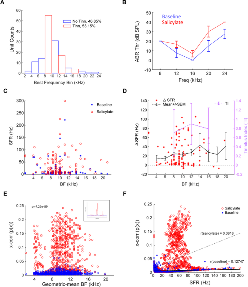
Figure 4. Salicylate induces increased ABR thresholds, increased SFR and synchrony in DCN fusiform cells.

A) Fraction of units by best frequency, with no-tinnitus frequencies (blue bars) and tinnitus frequencies (red bars) indicated. 46.85% of units were within a no-tinnitus band, while 53.15% of units were in a tinnitus band. B) ABRs were measured before surgery (blue) and post-salicylate administration after 30 minutes had past (red). C) Fusiform cells in all test animals showed significant increases in SFR after salicylate administration (red circles) compared to baseline (blue stars). D) Change in SFR for each unit from baseline to salicylate (red stars; mean+/− SEM indicated by dashed black line). Most increases occur in frequencies where GPIAS-measured tinnitus was confirmed (pink line). E) Cross-unit synchrony between pairs of spiketrains was computed at baseline (blue) and post-salicylate (red; see inset for sample cross-correlations) and is significantly increased over range of geometric mean BFs of sampled pairwise spiketrains (ANOVA2; p=7.26e-89, n=4404). F) Synchrony and SFR increase their correlation (r=0.3818) post-salicylate administration compared to baseline (r=0.127; Pearson’s linear correlation). Data shown are mean+/− SEM; alpha = 0.05.
