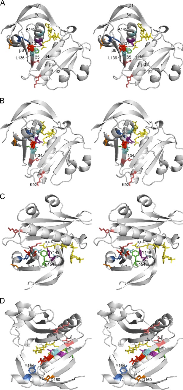
FIG 9.
Stereo views of B. anthracis SatA structure. Different views of the crystal structure of the SatA from Bacillus anthracis strain Ames (PBD entry 3PP9) are shown. Acetyl-CoA is shown in yellow, and residues discussed in the text are colored and labeled. (A) The β-sheets are labeled, showing the splaying of β4 and β5 to allow the acetyl-CoA to bind. E137 (red) is predicted to be the active-site glutamate. A change to L136 (cyan) is predicted to interfere with residue E137, while a substitution for residue A145 (cyan) is predicted to disrupt acetyl-CoA binding to SatA. (B) Residues I134 and K92 (salmon) are found at the edges of β-sheets. Substitutions for these residues may impact overall folding of the protein. (C) Residues Y149 (purple) and F154 (green) form a cavity around the acetyl group of acetyl-CoA, forming a putative active-site pocket. The hydroxyl group of Y149 is ~3.4 Å from the sulfur atom of acetyl-CoA and may act as a general acid to reprotonate the thiolate after transfer of the acetyl moiety. (D) Residue Y164 (blue) is parallel to the acetyl group of acetyl-CoA and E137 and is predicted to help position streptothricin for catalysis. Residue D160 (orange) is positioned on the dimer interface and is important for proper dimerization.

