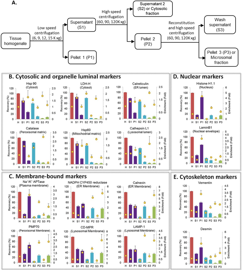
Figure 1. Recovery and enrichment of organelle markers in different subcellular fractions isolated during microsomal preparation.

A. Schematic figure of microsomal preparation by differential centrifugation. Recovery (bars) and enrichment (dots) of organelle markers (B. Cytosolic and organelle luminal markers; C. Membrane-bound markers; D. Nuclear markers and E. Cytoskeleton markers) in different liver subcellular fractions using typical low- (9,000 xg) and ultra- (120,000 xg) centrifugation speeds (mean±S.E., n=3) after homogenization with bead homogenizer (3 cycles). H: Homogenate; S1: S9 fraction; P1: Pellet 1 (heavy membrane); S2: Cytosolic fraction; P2: Pellet 2; S3: Wash supernatant; P3: Microsomal fraction.
