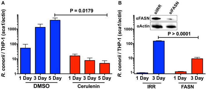
Figure 8.
Inhibition of FASN function and expression inhibits the ability of R. conorii to proliferate within THP-1 cells. (A) Quantitative PCR analysis of gDNA from PMA differentiated THP-1 cells infected with R. conorii Malish7 in the presence of DMSO (vehicle control) or FASN inhibitor, cerulenin, at 24 h, 3, and 5 days-post treatment. (B, Inset) Western immunoblot analysis confirms the inhibition of FASN protein expression from samples isolated at 3 days-post transfection when compared to control RNA treated cells (siIRR). An immunoblot for Actin is used to control for equal protein loading in each lane. Quantitative PCR analysis of gDNA from PMA differentiated THP-1 cells transfected with siRNA against FASN or a control RNA and infected with R. conorii Malish7 at an MOI of 2. Samples were analyzed at 24 h and 3 days post infection. Data is representative of two independent experiments with each condition in triplicate. Statistical analysis was performed by One-Way ANOVA with a Dunnett's post-hoc test for pairwise comparison. Statistical significance (p < 0.05).

