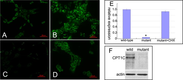
Figure 2.

The pathogenic investigations of mutant CPT1C. The GFP fluorescence showed a normal expression in 293T cells transfected with pEGFP‐N1 vectors (A), an increased expression in 293T cells transfected with wild‐type pEGFP‐N1‐CPT1C vectors (B), and a significantly decreased expression in 293T cells transfected with c.226C>T mutant pEGFP‐N1‐CPT1C vectors (C). After administration of cycloheximide (CHX), the expression of mutant CPT1C was recovered (D). Quantitative PCR measurements showed a significant reduction in the mutant CPT1C transcript expression compared to the level of wild‐type transcript. Repeated three times for every test. Data were analyzed by one‐way ANOVA test; Error bars are SEM; *P < 0.01(E). Immunoblot showed that no full‐length or truncated CPT1C proteins were detected in the mutant cells (F).
