
Figure 8.
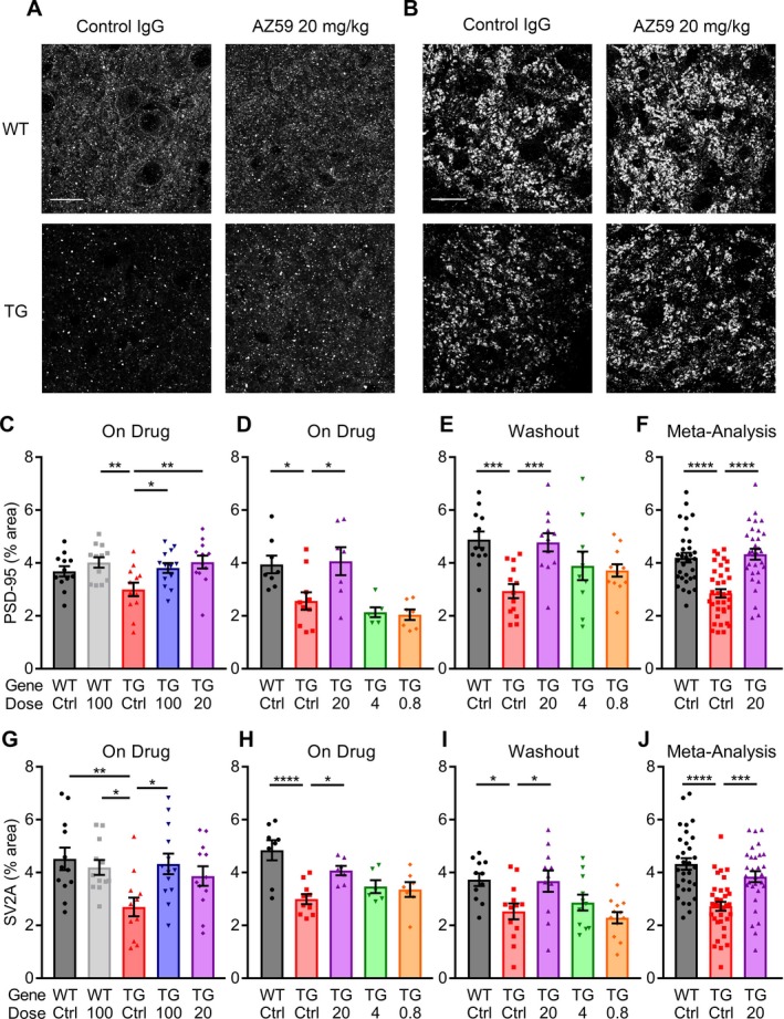
AZ59 reversal of synaptic deficits in APP/PS1 is dose‐dependent and persists after washout. (A–B) Representative images of PSD‐95 (A) and SV2A (B) taken in the dentate gyrus of 14‐month‐old WT and APP/PS1 mice after 5 weeks of treatment. Scale bar = 20 μm. (C–F) Quantification of PSD‐95 immunoreactive area in the dentate gyrus of treated mice. (C) 14‐month‐old mice after 5 weeks of treatment. (D) 12‐month‐old mice after 7 weeks of treatment. (E) 13‐month‐old mice after 7 weeks of treatment and a one‐month washout. (*, P < 0.05; **, P < 0.01; ***, P < 0.001) n = 6–13 per group. (F) Meta‐analysis of all PSD‐95 data for the WT control IgG, APP/PS1 control IgG, and APP/PS1 20 mg/kg AZ59. (****, P < 0.0001) n = 31–35 mice. All data are mean ± SEM, one‐way ANOVA with Dunnett's comparison to APP/PS1 control IgG. (G–J) Quantification of SV2A immunoreactivity in the dentate gyrus of treated mice. (C) 14‐month‐old mice after 5 weeks of treatment. (D) 12‐month‐old mice after 7 weeks of treatment. (E) 13‐month‐old mice after 7 weeks of treatment and a one‐month washout. (*, P < 0.05; **, P < 0.01; ****, P < 0.0001) n = 6–13 per group. (J) Meta‐analysis of all SV2A data for the WT control IgG, APP/PS1 control IgG, and APP/PS1 20 mg/kg AZ59. (***, P < 0.001; ****, P < 0.0001) n = 31–35 mice. All data are mean ± SEM, one‐way ANOVA with Dunnett's comparison to APP/PS1 control IgG.