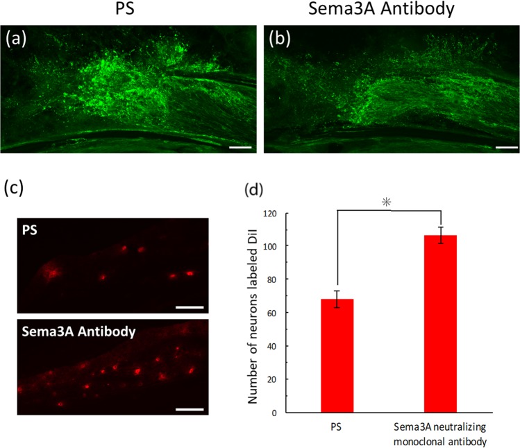
Figure 3.
Effects on IAN regeneration with a local administration of anti-Sema3A antibody. (a,b) Nerve regeneration demonstrated with PGP9.5 immunohistochemistry at postoperative (PO) 3 days. (a) Nerve fibers extending in random direction from the lesion are observed in a vehicle control treated with physiological saline (PS). In contrast, administration of anti-Sema3A antibody eliminates the radial extension of injured axons, resulting in axonal regeneration (b). (c) A comparison of DiI-labeled neurons in the trigeminal ganglion with a local administration of PS and Sema3A antibody to the transected site of IAN. The graph (d) shows that the number of DiI-labeled neurons is greater in anti-Sema3A antibody-treated animals than in vehicle control animals with a significant difference (Student’s t-test, *p < 0.05). Scale bars = 100 µm (a–c).

