Fig. 1.
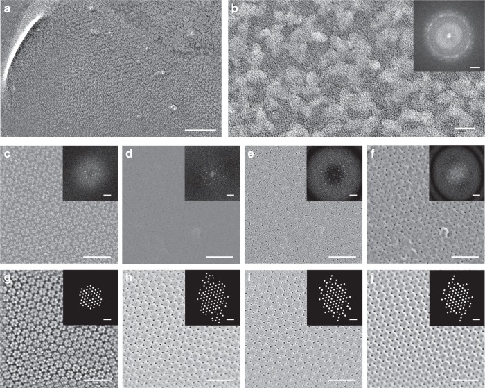
Platinum crystallization in conventional freeze-fracture replicas and resolution improvement of amorphous carbon replicas. a Electron micrograph of a conventional rotary shadowed freeze-etch of uroplakin complexes on the luminal surface of the bladder epithelium. b High magnification micrograph of a single uroplakin complex and its power spectrum (inset). c–f Direct comparison of uroplakin complex freeze-etch replicas using conventional rotary shadowing (c) and carbon shadowing imaged at near focus (d), defocus (e) or with a phase plate (f). Power spectra (insets) show the diffraction spots arising from uroplakin lattice. g–j The images in c–f were Fourier filtered to highlight the lattices in a conventional replica (g), and carbon replicas imaged near focus (h), defocus (i), or with a phase plate (j). The spot positions are highlighted in the spatial frequency mask (insets in g–j). These masks were used to generate the filtered images. Scale bars = 100 nm for a, 5 nm for b, 2 nm−1 for b inset, 50 nm for c–j, and 0.2 nm−1 for the power spectrum insets in c–j

