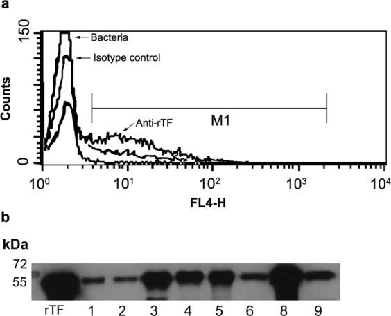
Figure 2.

CW localization of TF. (a) R6 bacteria were incubated with either: i. anti-rTF mAb or ii. isotype control mouse serum (as indicated) or iii. phosppate buffered seline. All were stained with Alexa Fluor 647®-conjugated goat-anti-mouse-IgG as a secondary antibody and then analyzed by flow cytometry. (b) Thirty micrograms of protein of CW fractions from 60S. pneumoniae clinical strains (see Supplementary Table S2) were loaded per lane, subjected to SDS-PAGE, and immunoblotted. Purified untagged rTF (0.01 μg) was loaded as a positive control. A representative blot is shown cropped from the 15 min exposed blot is presented.
