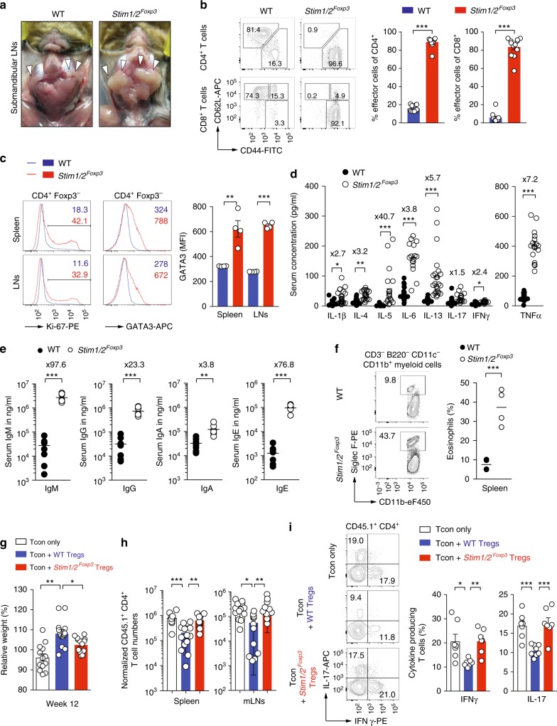
Fig. 2.
STIM1 and STIM2 control Treg function and prevent Th2-mediated autoimmunity. a Macroscopic analysis of submandibular LNs (arrows) in male WT and Stim1/2Foxp3 mice. b Analysis of naive (CD62LhiCD44lo), effector (CD62LloCD44hi), and memory (CD62LhiCD44hi) CD4+ and CD8+ T cells in the spleen of WT and Stim1/2Foxp3 mice by flow cytometry; means ± SEM of 10–12 mice. c Analysis of cell cycle (Ki-67) and GATA3 expression in conventional CD4+ T cells in the spleen and LNs of male WT and Stim1/2Foxp3 mice by flow cytometry; means ± SEM of four mice. d ELISA measurements of serum cytokines in male WT and Stim1/2Foxp3 mice; fold increases in Stim1/2Foxp3 mice compared to WT mice are indicated by ×; means ± SEM of 13–20 mice. e Analysis of serum immunoglobulin concentrations in male WT and Stim1/2Foxp3 mice using ELISA; means of eight mice. f Analysis of CD11b+Siglec F+ eosinophils in the spleen of male WT and Stim1/2Foxp3 mice by flow cytometry; means of four mice. g–i Stim1/2-deficient Treg cells fail to suppress adoptive transfer colitis in lymphopenic host mice; for details see Supplementary Figure 2d. g Weight of Rag1–/– host mice 12 weeks after transfer of CD45.1+ conventional CD4+ WT T cells together with CD45.2+ WT or Stim1/2-deficient Treg cells isolated from female heterozygous Stim1/2Foxp3 mice; means ± SEM of 13–14 host mice. h Normalized numbers of CD45.1+ conventional CD4+ T cells in spleen and mLNs of Rag1–/– host mice 12 weeks after transfer; means ± SEM of 7–8 host mice. i Analysis of IL-17A and IFNγ production by CD45.1+ conventional T cells from mLNs after PMA/ionomycin re-stimulation using flow cytometry; means ± SEM of 7–8 host mice. Statistical analysis in b–g and i by unpaired Student’s t-test, in (h) by one-way ANOVA. *p < 0.05; **p < 0.01; ***p < 0.001

