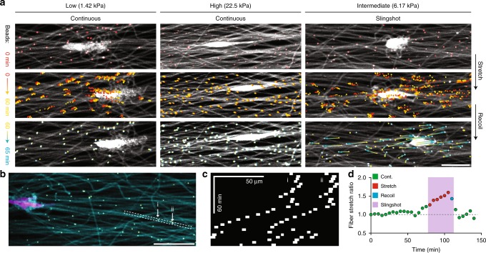
Fig. 4.
Slingshot migration involves stretch and recoil of matrix fibers. a Representative time-lapse images of fibers containing fluorescent microspheres, used as fiducial markers to examine matrix deformations underlying continuous and slingshot migration (scale bar: 50 µm). b Composite confocal fluorescence image of an NIH3T3 within an intermediate stiffness matrix (matrix fibers (cyan), cytoplasm (magenta), and fiber-embedded beads (yellow); scale bar: 50 μm). c Kymograph of a pair of microspheres embedded within the same fiber, as indicated in b used to determine fiber stretch ratio (d) (relative to initial distance between beads) as a function of time

