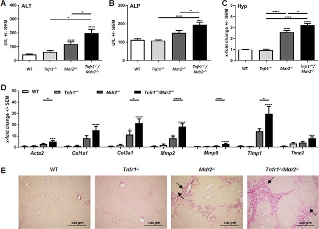
Figure 1.
Absence of TNFR1 increased tissue injury in the Mdr2−/− mouse model. (A) ALT and (B) ALP levels determined in plasma of WT (n ≥ 4), Tnfr1−/− (n ≥ 6), Mdr2−/− (n ≥ 9), and Tnfr1−/−/Mdr2−/− (n ≥ 9) mice. (C) Quantification of the hepatic hydroxyproline content of mice described in A. (D) Relative hepatic expression of Acta2, Col1a1, Col3a1, Mmp2, Mmp9, Timp1, and Timp2 of mice described in A, determined by RT-qPCR. (E) Representative images (10x) of Sirius Red stained tissue sections of mice described in (A). *P ≤ 0.05, ***P ≤ 0.001, ****P ≤ 0.0001.

