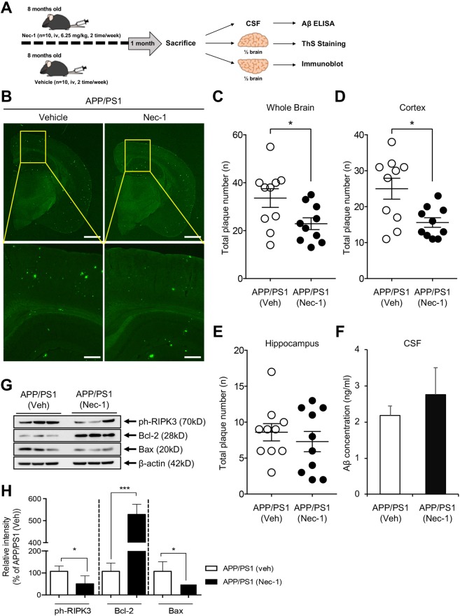
Figure 4.
Nec-1 reduces Aβ plaques in aged APP/PS1 mouse brains. (A) Schedule of Nec-1 administration. Nec-1 (6.25 mg/kg, n = 10) or vehicle (2.5% DMSO in PBS, n = 10) was injected into 8-month-old male APP/PS1 mice via tail vein for 4 weeks (2 times per week). Brains and CSF samples were collected after sacrifice. The illustration was drawn using Adobe Photoshop software program. (B) ThS-stained Aβ plaques in whole brains of APP/PS1 mice treated with vehicle (n = 10) or Nec-1 (n = 10). Data presented in this article are 4 representative images. Scale bars = 1 mm (upper), 200 μm (lower). Total numbers of ThS-positive Aβ plaques in whole brain (C), cortex (D), and hippocampus (E) of APP/PS1 mice after Nec-1 administration. Numbers of plaques were reduced by Nec-1 administration. (F) Aβ42 levels in cerebrospinal fluid from five mice which used for ThS staining in aforementioned mice with or without Nec-1 treatment. (G) Western blot analysis and (H) quantification of phosphorylated RIPK3, Bcl-2, and Bax to observe changes of necroptosis and apoptosis. Three mice from each group were analyzed for this experiment. Phosphorylated RIPK3 and Bax were reduced, while Bcl-2 was increased by Nec-1 administration. Data is presented as mean ± SEM. *P ≤ 0.05 and ***P ≤ 0.001 (One-way ANOVA followed by Bonferroni post-hoc comparison tests). Full-length original blots are shown in Supplementary Information.

