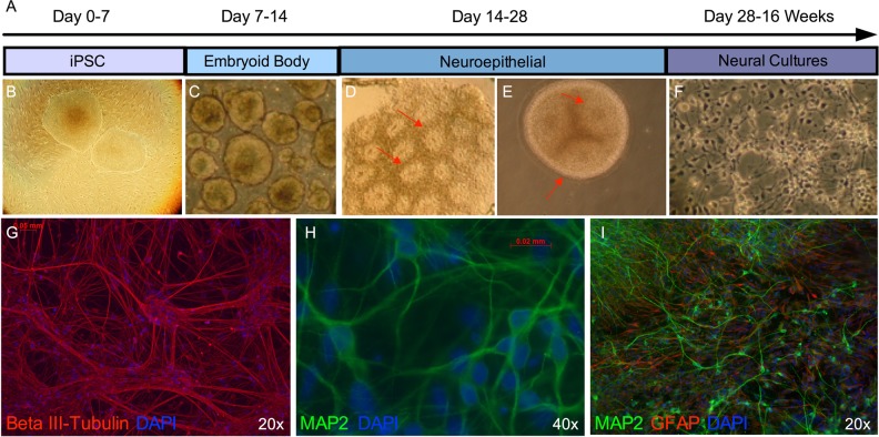
Fig. 1.
Neural differentiation of human iPSCs. a Schematic depicting the neural differentiation protocol. Induced pluripotent stem cells (b) are cultured on irradiated mouse embryonic fibroblasts for 7 days, following which they are cultured in suspension for 7 days to allow for the formation of embryoid bodies (c). Embryoid bodies are plated on a laminin substrate for 7 days to generate neuroepithelial cells (d), which form neural rosette-like structures (indicated by red arrows). Neuroepithelial cells are cultured in suspension for an additional 7 days to form and expand neurospheres (e), which display neural rosette-like structures (red arrows), before being manually dissociated and plated onto glass coverslips in neural media (f). After 12 weeks in neural media, cultures contain numerous Beta III-tubulin-positive neurites (g), pyramidal shaped MAP2-postive neurons (h), and GFAP-positive astrocytes (i)

