Figure 1.
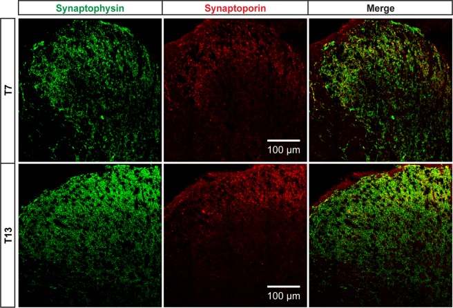
Central distributions of synaptophysin and synaptoporin in the T7 and T13 spinal cord dorsal horn in rats. Immunohistochemistry for presynaptic vesicle proteins, synaptophysin (green) and synaptoporin (red), shows that synaptophysin is distributed across dorsal horn laminae whereas synaptoporin is localized in superficial laminae I-II. Double-labeling of synaptic terminals positive for both synaptophysin and synaptoporin is visualized as yellow in merged images. Shown are collages of 16 confocal images at 40X objective lens.

