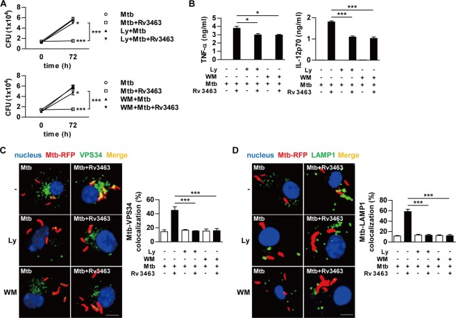
Figure 6.
PI3K inhibition interferes the Rv3463-mediated activities. Bone marrow-derived macrophages (BMDMs) pretreated with pharmacological inhibitors of PI3K (20 μM LY2940029 [Ly] or 200 nM Wortmannin [WM]) for 1 h were infected with Mtb (A,B) or Mtb-RFP (C,D) at a multiplicity of infection of 1 for 4 h, treated with amikacin, washed, and incubated with or without 5 μg/ml Rv3463 for 72 h. (A) Intracellular Mtb growth in the cells was determined at 0 and 72 h. *p < 0.05 and ***p < 0.001 for treatment compared to infection only control. (B) TNF-α or IL-12p70 production in culture supernatants were measured by ELISA. (C,D) Colocalization of VPS34 or Lamp1 molecules (green) with Mtb (red) in the treated BMDMs were analyzed by laser-scanning confocal microscopy. The cells were stained with DAPI to visualize the nuclei (blue). Scale bar, 10 μm. Bar graphs show the quantification of VPS34 or Lamp1 colocalization with the Mtb phagosome. Values are mean ± SD of 50–100 cells per each experiment (n = 3). *p < 0.05, **p < 0.01 and ***p < 0.001 for inhibitor treatment compared to the Rv3463-treated cells.

