
Figure 7.
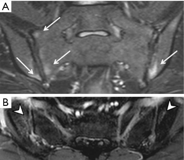
Sacroiliitis on MRI. (A) Bone marrow oedema (arrows) on T2W FS oblique coronal images and (B) corresponding enhancement (arrowheads) on T1W FS post-contrast axial images. This degree of bone marrow oedema is sufficient to diagnose sacroiliitis.