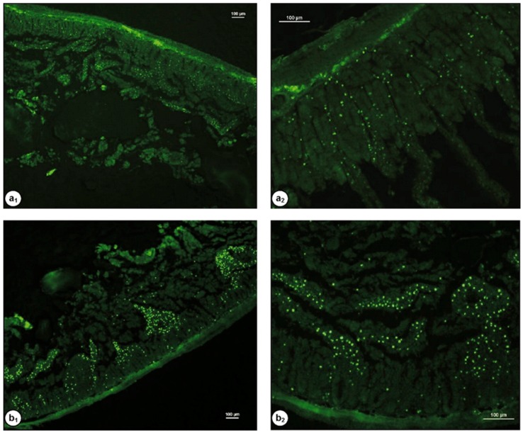
Figure 9.
Fluorescent images of rat intestinal tissues after 2 h incubation with 100 µL (0.5% w/v) chitosan (a) and chitosan-TBA (b) nanoparticles labelled with Alexa Fluor 488, (a1 and b1, 40×; a2 and b2, 100× magnification). The scale bars = 100 µm. Reprinted from [99] with permission of Elsevier.

