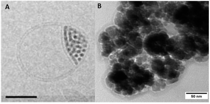
Figure 7.
Cryo-TEM (transmission electron microscopy) micrographs of (A) lipid bilayer splitting around incorporated MNPs (scale bar = 50 nm). Reprinted from [151] with permission of ACS Publications. (B) Protein corona of bovine serum albumin formed at the surface of MNPs (unpublished own data).

