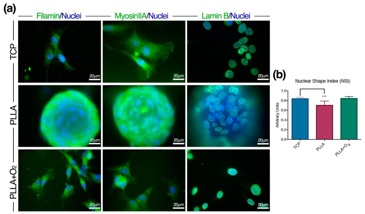
Figure 4.
hASCs architecture on PLLA and on PLLA+O2. (a) Representative immunofluorescence images of Filamin-A, Myosin-IIA and Lamin-B proteins in hASC spheroids on PLLA, and fibroblast-like hASCs on PLLA+O2 and on TCP; and (b) NSI in hASCs on PLLA, PLLA+O2, and TCP (see methods for details). *** p ≤ 0.001.

