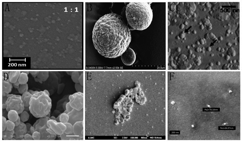
Figure 4.
Scanning electron microscopy (SEM) or scanning force microscopy (SFM) micrographs of synthesized chitosan nanoparticles using six different techniques. (A) SEM micrograph of the nanoparticles modified through emulsion crosslinking. Reproduced with permission from [61]; (B) SEM micrograph of the nerve growth factor-loaded chitosan nanoparticles modified through ionically crosslinking. Reproduced with permission from [62]; (C) SFM micrograph of the chitosan-modified (poly(d,l-lactide-co-glycolide); PLGA) nanoparticles modified through solvent evaporation. Reproduced with permission from [63]; (D) SEM micrograph of the cellulose-chitosan complex nanoparticles modified through spray drying. Reproduced with permission from [64]; (E) SEM micrograph of the nanoparticles modified through precipitation, reproduced with permission from [66]; (F) SEM micrograph of the chitosan-alginate nanoparticles modified through chitosan solution coating. Reproduced with permission from [67].

