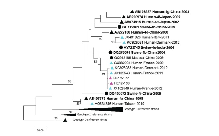
Figure 3.
Phylogenetic analysis of hepatitis E virus genotype 4 isolates, Belgium, 2010–2017 (n = 14 human sequences)
HEV: hepatitis E virus.
Maximum-likelihood phylogenetic tree of a 348 bp fragment from open reading frame 2. Genetic distances were calculated using the Tamura Nei model. Only bootstrap values > 70 are reported. GenBank accession numbers are shown for each HEV reference strain used in the phylogenetic analysis and are noted as follows: Accession number_Host-HEV subtype-Country-Year. Triangles represent human HEV sequences and circles animal sequences.
Rose symbols: 2012 Belgian HEV-4 sequences; black symbols: HEV reference strains according to Smith et al. [17]; light blue symbols: other human and animal HEV sequences.

