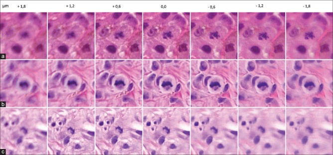
Figure 1.
Examples of changes in the status of dermal mitotic activity by pathologist 1 after the use of software focusing. Case 7 (a) changed into true positive. Case 59 (b) changed into false negative. Case 97 (c) changed into true positive. Note that the best field of view of the mitosis is situated mainly below and above the zero z-plane in cases 7 and 97, respectively

