Figure 4.
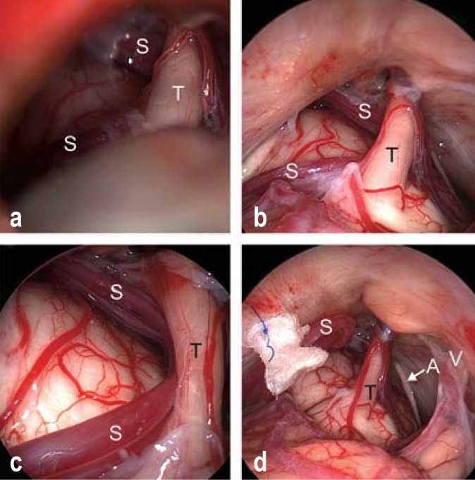
Endoscopically assisted microvascular decompression for trigeminal neuralgia.
a) View of the trigeminal nerve (T) and the superior cerebellar artery (S) through the operating microscope.
b) Inspection through the endoscope (0° optics), revealing the entire cisternal course of the trigeminal nerve (T) and the superior cerebellar artery (S).
c) Inspection through the endoscope with 30° optics reveals severe vascular compression of the trigeminal nerve (T) by the elongated loop of the superior cerebellar artery (S).
d) The superior cerebellar artery (S) has been transposed and sewn upward toward the tentorium with a loop of Teflon. Ideally, the trigeminal nerve (T) should be entirely free after the decompression, without any contact either with the vessel or with the Teflon pledget. The abducens nerve (arrow A) and the vestibulocochlear nerve (V) can be seen in the vicinity.

