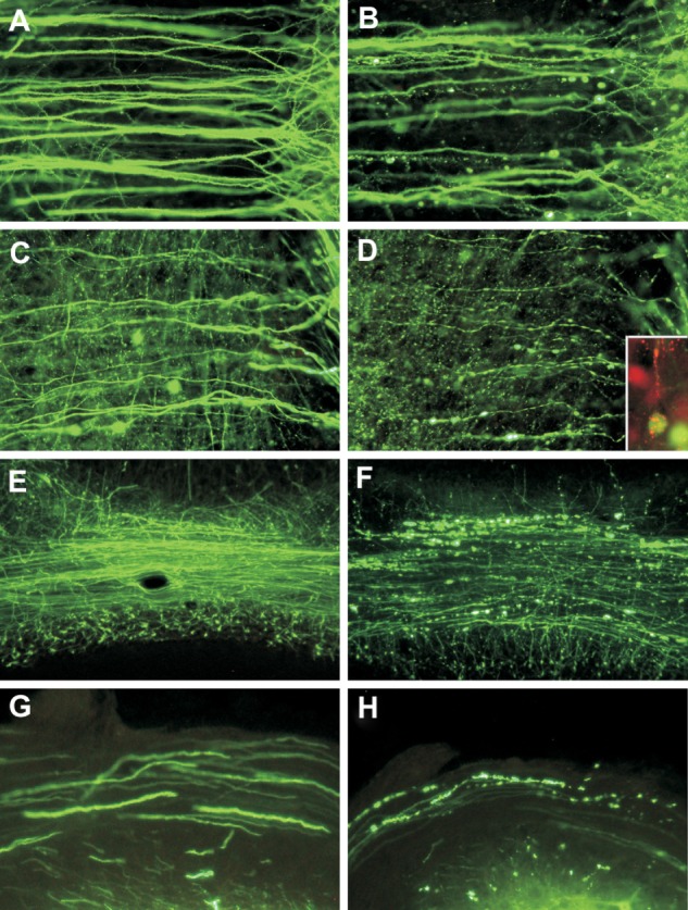
Figure 1.

Fluorescence microscopy showing dendrites (A and B) and axons (C and D) of layer V pyramidal neurons in the cerebral cortex of mock-infected (A and C) and moribund challenge virus standard-infected (B, D, and D inset) yellow fluorescent protein mice.43 In infected mice, beading is observed in a minority of dendrites (B), and more axons are involved (D). There are no abnormalities in the dendrites (A) or axons (C) of mock-infected mice. Axons in mock-infected mice are slightly varicosed (C), which is characteristic of these fibers. Fluorescence microscopy shows rabies virus antigen (red) in the perikaryon and dendrite of a yellow fluorescent protein-expressing neuron (D inset). Morphology of the cerebellar mossy fibers of mock-infected (E) and moribund challenge virus standard-infected yellow fluorescent protein mice (F). Mossy fiber axons in the cerebellar commissure of moribund mice show severe beading (F), whereas no abnormalities were observed in mock-infected mice (E). Axons in the inferior cerebellar peduncles are normal in mock-infected mice (G) and show marked beading in challenge virus standard-infected moribund mice (H). A–D) ×230; D) inset, ×220; E, F) ×80; G, H) ×350.
Copyright © 2010, American Society for Microbiology. Adapted with permission from Scott CA, Rossiter JP, Andrew RD, Jackson AC. Structural abnormalities in neurons are sufficient to explain the clinical disease and fatal outcome in experimental rabies in yellow fluorescent protein-expressing transgenic mice. J Virol. 2008;82:513–521.43
