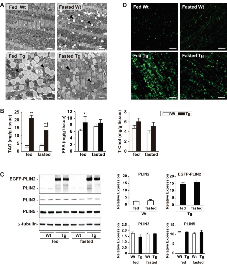
Fig. 3.
Effect of fasting on PLIN2-induced cardiac steatosis. A: representative electron micrographs showing LD accumulation in the left ventricles of WT and Tg mice under feeding and 24-h-fasting conditions. Note the accumulation of small LDs in WT mice (arrowheads) after 24 h of fasting (top right) compared with smaller and “shrunken” LDs after 24 h of fasting in Tg hearts (bottom). Scale bars, 2 μm. B: cardiac contents of TAG, free fatty acid (FFA), and total cholesterol (T-Chol) in WT and Tg mice under feeding and 24-h-fasting conditions. Values are means ± SE of 4–6 mice in each group. *P < 0.05 and **P < 0.01 vs. WT mice; †P < 0.05 vs. feeding condition. C: Western blot analysis of PLIN2, PLIN3, and PLIN5 in ventricles of WT and Tg mice under feeding and 24-h-fasting conditions. Anti-PLIN2, -PLIN3, -PLIN5, and -α-tubulin antibodies were used in these experiments. Six mice per group were analyzed, and representative blots are presented. Graphs: densitometric analysis of the protein expression normalized by α-tubulin. Values are means ± SE of 4 mice/group. D: PLIN2 expression in cardiomyocytes of WT and Tg mice under feeding and 24-h-fasting conditions. The tissue sections were stained with anti-PLIN2 antibody and analyzed by confocal microscopy. Four mice in each group were studied, and representative images are shown. Scale bars, 10 μm.

