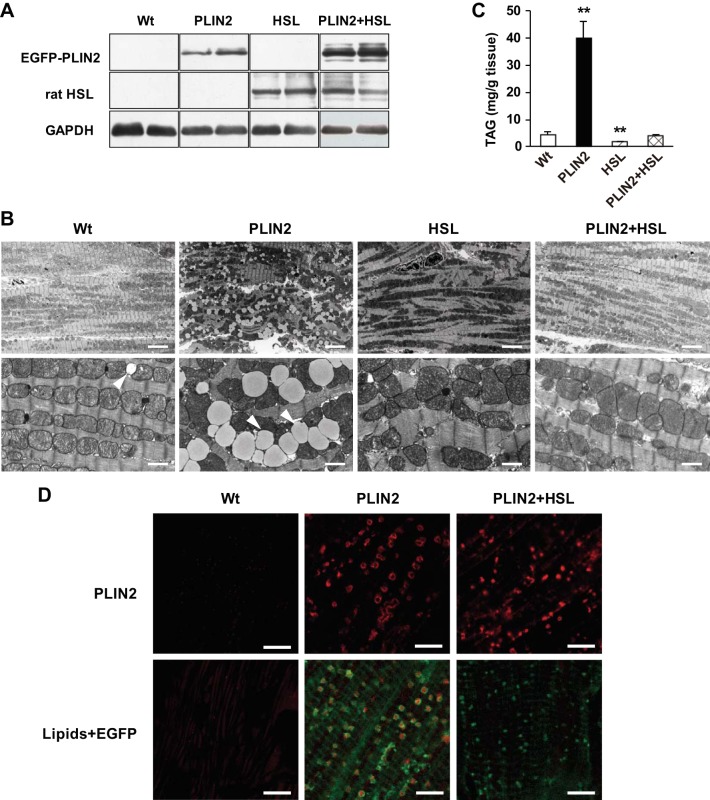
Fig. 7.
Effects of HSL overexpression on PLIN2-induced cardiac steatosis in HSL-PLIN2 double-Tg mice. A: Western blot analysis of EGFP-PLIN2, rat HSL, and GAPDH using specific antibodies against each protein. Representative data of 4–5 mice/group. PLIN2, PLIN2-Tg mice; HSL, HSL-Tg mice; PLIN2 + HSL, PLIN2-HSL double-Tg mice. B: electron micrographs of the left ventricles. Arrowheads, LDs. Scale bars, 6.7 (top) or 1 μm (bottom). C: cardiac TAG content in WT and Tg mice. Tissue lipids were extracted with chloroform-methanol, and TAG contents were measured using a TAG assay kit. Values are means ± SE of 4–5 mice/group. **P < 0.01 vs. WT mice. D: PLIN2 expression and LD accumulation in left ventricles from WT, PLIN2-Tg, and PLIN2-HSL double-Tg mice. The tissue sections were stained with anti-PLIN2 antibody (top) or LipidTOX (bottom) and analyzed by confocal microscopy. At the bottom are merged images of those focused on LDs (red) and EGFP (green). Four mice in each group were studied, and representative micrographs are presented. Scale bars, 5 μm.

