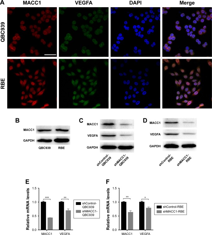
Figure 5.
Localization of MACC1 and VEGFA in human CCA cell lines and downregulation of VEGFA expression by MACC1 knockdown in CCA cells.
Notes: (A) Confocal laser scanning microscopy was used to determine the localizations of MACC1 (red) and VEGFA (green) in QBC939 cells (up) and RBE cells (down). Scale bars =50 µm. (B) MACC1 protein levels in QBC939 and RBE cells. (C, D) Impact of MACC1 knockdown on VEGFA levels in CCA cells assessed by Western blotting analysis. GAPDH was used as an internal control. (E, F) Impact of MACC1 knockdown on VEGFA levels in CCA cells assessed by real-time qPCR analysis. The assay was performed in three independent experiments. The expression levels were normalized to those of GAPDH. *P<0.05, **P<0.01, ***P<0.001.
Abbreviation: CCA, cholangiocarcinoma.

