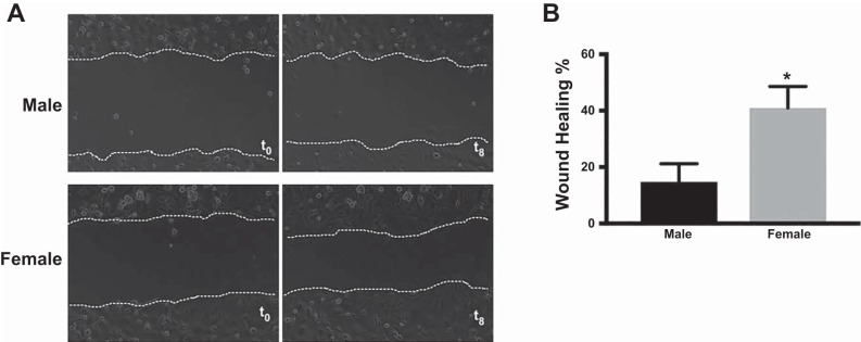
Fig. 1.
Two-dimensional wound healing assay reveals increased migration in female human pulmonary microvascular endothelial cells (HPMECs). A: representative images of male and female HPMECs taken at baseline [time 0 (t0)] and after 8 h incubation. B: percentage of wound closure at 8 h. Values are means ± SE of 3 independent biological replicates (n = 3/group). Significant differences between male and female HPMECs are indicated by *P < 0.05.

