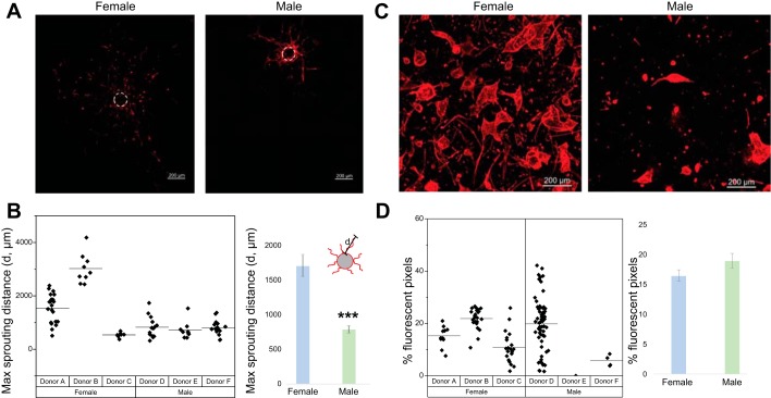
Fig. 2.
Female human pulmonary microvascular endothelial cells (HPMECs) exhibit greater sprouting angiogenesis. Angiogenic potential was quantified based on the maximum length of sprouts protruding from HPMEC-coated cytodex-3 microcarrier beads suspended in a fibrin gel. A: representative images from male and female HPMECs subjected to a sprouting angiogenesis assay. B: maximum sprouting distance in male and female HPMECs. The ability of cells to form a three-dimensional self-assembled vascular network, known as a plexus, was evaluated within collagen gels cultured over 7 days. Each point represents one coated bead. C: representative images from male and female HPMECs subjected to a vasculogenesis assay. D: percentage of fluorescent pixels (fluorescent pixels/total pixels). Each point represents one collagen gel. Values are means ± SE of 3 independent biological replicates (n = 3/group). Significant differences between male and female HPMECs are indicated by ***P < 0.001.

