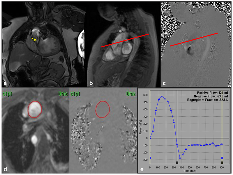
Fig. 2.
a Steady-state free precession (SSFP) left ventricular outflow tract (LVOT) cine imaging of a quadricuspid aortic valve with severe aortic regurgitation (yellow arrow). b, c “In-plane” phase-contrast velocity encoded imaging. The in-plane image allows for perpendicular planning (red line) to obtain through-plane images above the aortic valve leaflets. d Phase-contrast velocity encoded through plane images with a region of interest drawn in the aorta. e Plotted graph of forward flow and regurgitant flow with calculated regurgitant fraction of 52.8% and regurgitant volume of 63.9 mL indicating severe aortic regurgitation

