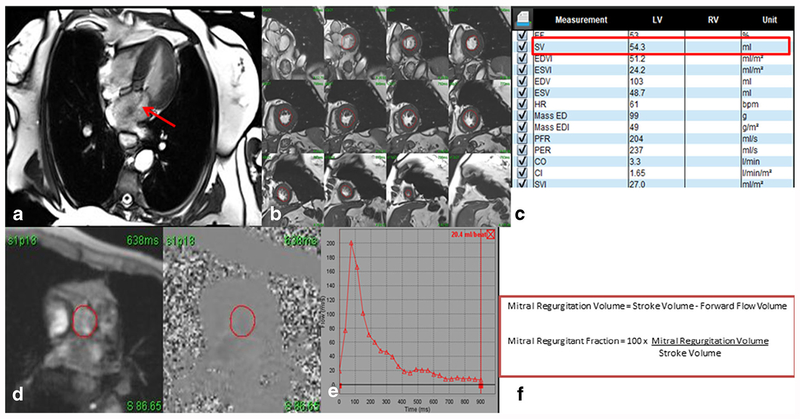
Fig. 3.
Four-chamber steady-state free precession (SSFP) cine image showing mitral regurgitation (red arrow) (a). Quantification was performed using volumetric analysis by obtaining short axis cine stacks (b) to obtain a stroke volume (c). Velocity encoded imaging of the aorta (d) above the level of the valve is obtained for forward-flow volume measurement (e). The regurgitant fraction can be obtained with a simple mathematical formula using the stroke volume and the forward flow volume (f). In this case, the regurgitant fraction of 62% indicates severe mitral regurgitation

