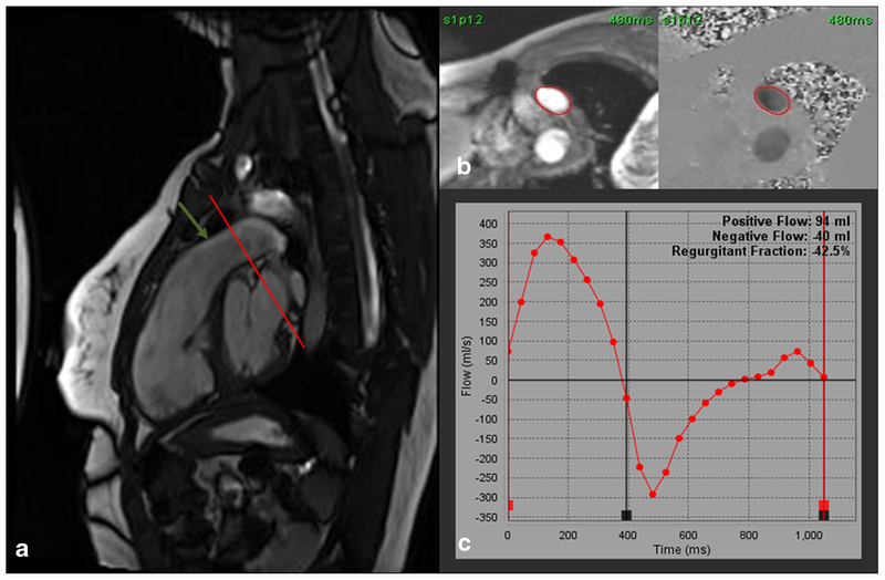
Fig. 4.
a Steady-state free precession (SSFP) right ventricular outflow tract (RVOT) cine image showing severe pulmonic regurgitation. The regurgitation is so severe that there is no turbulent flow to cause dephasing and a low signal flow void. A through-plane phase-contrast velocity-encoded image (VENC) can be obtained at the cross-sectional plane (red line) of the pulmonary artery (green arrow). b Through-plane VENC image with a region of interest drawn in the pulmonary artery. c Graphic plot of forward and regurgitant flow through the pulmonary artery showing a regurgitant fraction of 42.5% indicating severe pulmonic regurgitation

