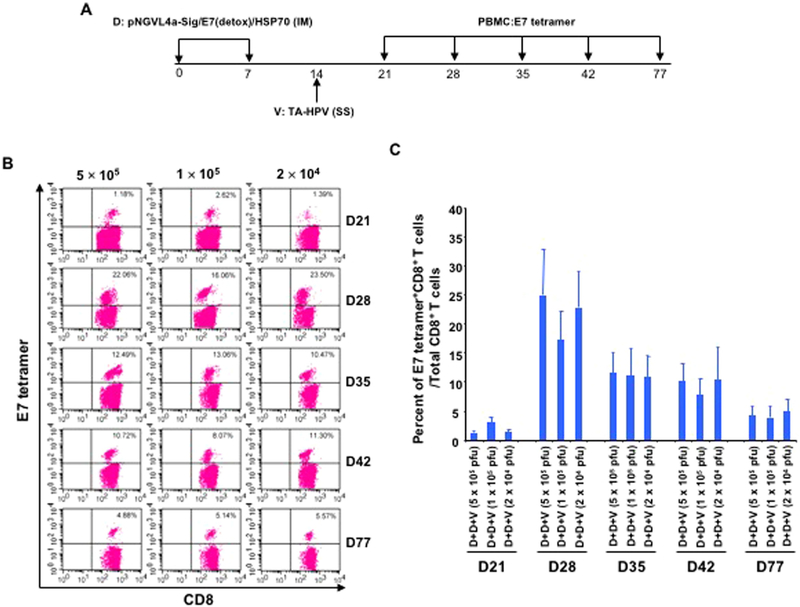
Fig. 4. Response of HPV16 E7-specific CD8+ T cells to different doses of TA-HPV administered through skin scarification.
A. Schematic illustration of the experiment. Briefly, groups of 5–8-week old female C57BL/6 mice (5 mice/group) were vaccinated with 25 μg/mouse of pNGVL4a-Sig/E7(detox)/HSP70 DNA vaccine in 50 μL via IM injection (hind leg muscle). The mice were boosted with the same regimen 7 days later. One week after the last vaccination, the mice were further boosted with either 5 × 10⁵, 1 × 10⁵, or 2 × 10⁴ pfu of TA-HPV (5 μL) on the tail through SS. 7, 14, 21, 28 and 63 days after the last vaccination, PBMCs were prepared and stained with anti-mouse CD8 and HPV16/E7 tetramer. The data were acquired with FACSCalibur flow cytometer and analyzed with CellQuest. B. Representative flow cytometry data from each group. C. Summary of the flow cytometry data.

