
Figure 4.
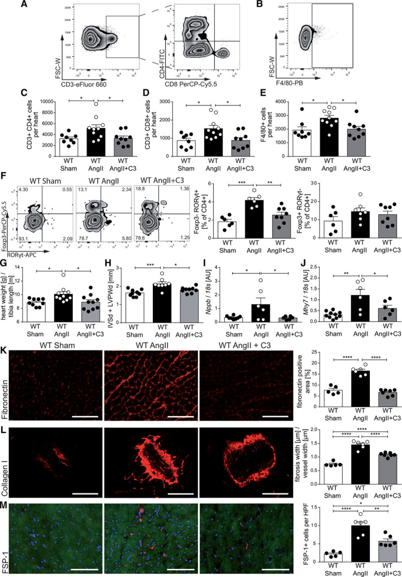
Propionate attenuates hypertensive cardiac damage in AngII-infused wild-type NMRI (WT) mice. A through E, Single cells were isolated from hearts of sham-infused or AngII-infused WT mice treated with C3 or control and analyzed by flow cytometry for T helper cells (CD3+CD4+), cytotoxic T cells (CD3+CD8+), and macrophages (F4/80+), as well. A and B, Representative ratings. C through E, The respective quantifications. WT Sham n=8, WT AngII n=10, WT AngII+C3 n=9. F, Analysis of CD4+FoxP3+ and CD4+RORγt+ cells in heart single-cell suspensions. Left, Representative flow cytometry plots. Right, Quantifications. WT Sham n=6, WT AngII n=6 to 7, WT AngII+C3 n=8. G, Cardiac hypertrophy index (heart weight [g]/tibia length [m]), (WT Sham n=9, WT AngII n=10, WT AngII+C3 n=10). H, Left ventricular wall thickness (sum of IVSd and LVPWd) as measured by echocardiography (WT Sham n=9, WT AngII n=8, WT AngII+C3 n=9). Cardiac Nppb (I) and Mhy7 (J) expression as measured by qPCR at the end of the treatment (WT Sham n=10, WT AngII n=6, WT AngII+C3 n=6). K through M, Immunofluorescence analysis of cardiac left ventricular fibrosis using fibronectin (K), collagen I (L), and FSP-1 (M) antibodies (WT Sham n=5, WT AngII n=6, WT AngII+C3 n=7). Left, Representative photomicrographs (scale bar=100 µm). Right, Quantifications. *P<0.05, **P<0.01, ***P<0.001, ****P<0.0001 by 1-way ANOVA and Tukey post hoc. AngII indicates angiotensin II; APC, Allophycocyanin; AU, arbitrary unit; C3, propionate; FITC, fluorescein isothiocyanate; FSC-W, forward scatter width; FSP-1, fibroblast-specific protein 1; HPF, high-power field; IVSd, interventricular septal thickness at diastole; LVPWd, left ventricular posterior wall end diastole; PB, Pacific Blue; PerCP-Cy5.5, Peridinin-chlorophyll protein cyanine 5.5; and qPCR, quantitative polymerase chain reaction.