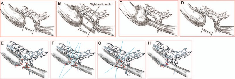
Figure 2.

Schematic diagrams of gross relative positions taking their orientation of the EA/TEF in the 4 cases. (A) Esophageal atresia with a distal tracheoesophageal fistula, the most frequently encountered form of esophageal anomaly. (B) Atresia appearance seemed with a double (proximal and distal) fistula on gross, but the esophageal atresia was confirmed only with a distal tracheoesophageal fistula and a right-sided aortic arch be found in operation. (C) Appearance seemed with H-type fistula but the double ends were not connected and there was only distal tracheoesophageal fistula with atresia. (D) The intra-operative pathologic anatomy showed esophageal atresia with a distal tracheoesophageal fistula. (E–H) Schematic pictures of the intra-operative “diamond-shape” anastomosis of EA performed for primary repair in the 4 cases. The esophageal atresia proximal blind end with a transverse cut and distal end with longitudinal cut, to make 2 esophageal end surface in “diamond-shape”. The absorb suture for 1-1’, 2-2’, 3-3’ and 4-4’ point to point corresponding anastomosis. Complete the “diamond-shape” anastomosis of EA. EA: Esophageal atresia; TEF: Tracheoesophageal fistula.
