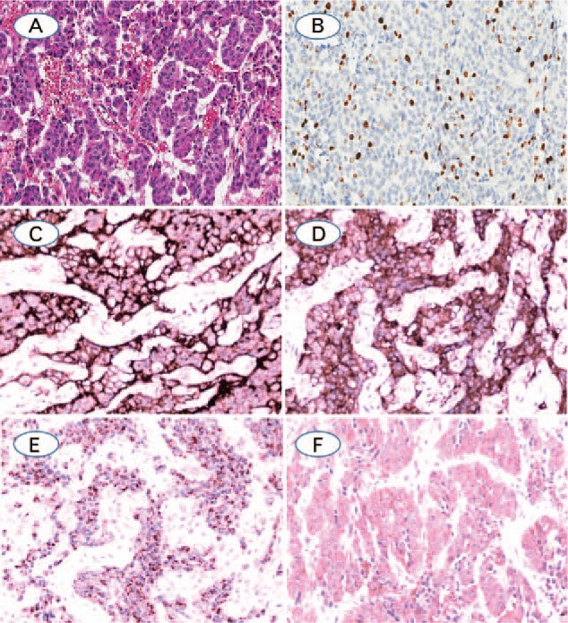
Figure 1.

(A) Typical carcinoid, with trabecular pattern with delicate intervening vascular stroma, no mitoses or necrosis are identified; (B) Ki-67, showing 12.66% positivity by computer image analysis and 15% positivity by manual counting in the hot spot; (C–E). TC showed diffuse positive expression for CD56, synaptophysin, CgA and MAP2, respectively (A: Hematoxylin-eosin staining; B–E: Immunohistochemistry staining. The original magnification of A, B was 100×, magnification for remaining cases were 200×).
