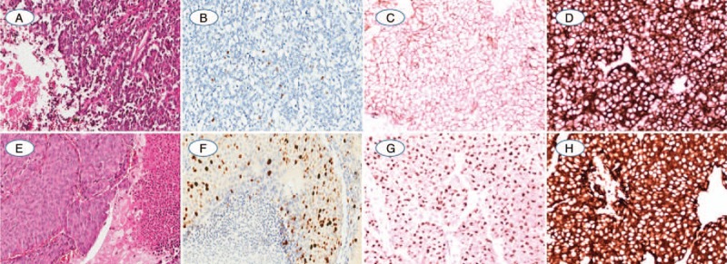
Figure 2.

(A) One AC showed vascularized stroma and spotty necrosis in the peripheral tumor clusters; (B) The Ki-67 index was evaluated as 4.34% positivity by CIAM and 2% positivity by MCM; (C, D). AC was diffuse strong positive expression of CD56 and synaptophysin; (E) Another AC with kindly cell morphology, focal necrosis, and 6 mitosis per mm2; (F) Ki-67 index was counted as 29.48% by CIAM and 25% by MCM with weak to strong intensity; (G, H). The neuroendocrine marker of CgA and Synaptophysin were diffuse positive expression in cytoplasm (A and E: Hematoxylin-eosin staining; B–D and F–H: Immunohistochemistry staining. The original magnification of A, B and E, F was 100×, magnification for remaining cases were 200×).
