
Figure 3.
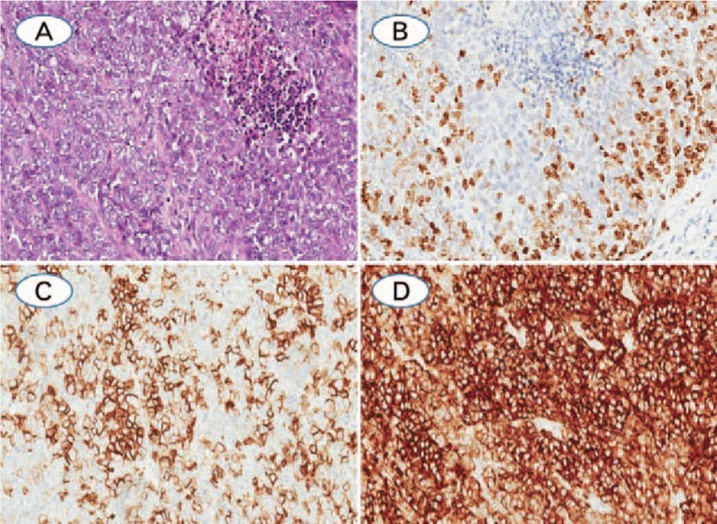
(A) Large cell neuroendocrine carcinoma with solid patterns, and the tumor cells are large, abundant cytoplasma, prominent nucleoli, and necrosis was observed; (B) Ki-67 index, which was evaluated as 54.48% by CIAM and 40% by MCM; (C) LCNEC was positive staining with the neuroendocrine markers of CD56; (D) Synaptophysin was diffuse positive expression in the cytoplasm (A: Hematoxylin-eosin staining; B–D: Immunohistochemistry staining. The original magnification of all cases were 200×).