Fig. 1.

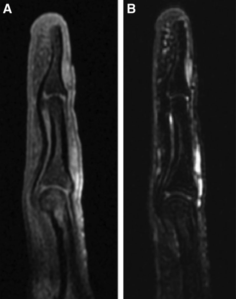
Preoperative magnetic resonance image showing the lesion at the subungual region of the right ring finger. A, T1-weighted image. B, T2-weighted image.

Preoperative magnetic resonance image showing the lesion at the subungual region of the right ring finger. A, T1-weighted image. B, T2-weighted image.