
Fig. 2.
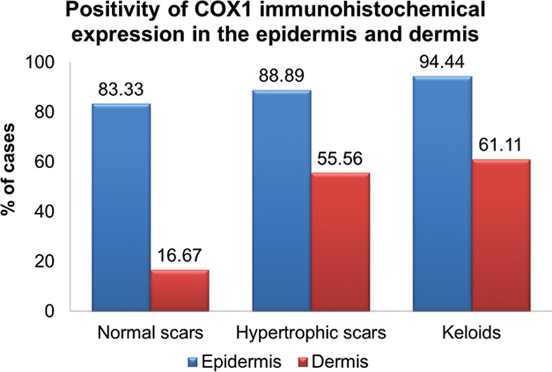
The graph shows the percentage of cases in each group in which the IHC expression of COX1 was positive. It is observed that, in the dermis there were more positive cases for COX1 in the groups of the hypertrophic and keloid scars compared with the group of normal scars.