Figure 1.
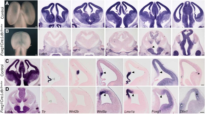
Early deletion of Ldb1 using Foxg1Cre results in reduced telencephalic size and disrupted telencephalic midline patterning. A, B, Whole brains and coronal sections at E12.5 from control (A) and Foxg1Cre;Ldb1lox/lox mutants (B) reveal a greatly reduced telencephalon on loss of Ldb1. Ldb1 is expressed in the entire control telencephalon and thalamus at E12.5 (A) but is undetectable in the mutant telencephalon (B). The expression boundaries of Ldb1 in the retina (open arrowhead, B) and the diencephalon (black arrowhead, B) are consistent with the reported activity of Foxg1Cre (Hebert and McConnell, 2000). C, D, E12.5 sections from control (C) and Foxg1Cre;Ldb1lox/lox mutant (D) brains. The expression of choroid plexus marker Ttr is lost in the mutant (open arrowheads, C, D). The mutant does not express hem marker Wnt2b but displays an expanded expression of Wnt5a and Lmx1a. Lmx1a labels both hem and choroid plexus in control sections. Foxg1 is expressed in a manner complementary to Lmx1a in control and mutant sections. Black arrowheads mark the cortex-hem boundary (C, D). Dbx1 is expressed in the septum (white asterisks) and antihem (black asterisks), both of which are expanded on loss of LDB1 (C, D). Scale bars: 100 μm.

