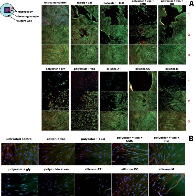
Figure 4.
(A) Global impact of the non-adhering dressings on fibroblast morphology and structure after 72 hours of direct contact with the dressing samples at the positions 1 – directly underneath the dressing sample, 2 – at the margin of the dressing sample applied and the surrounding cell layer, and 3 – from the surrounding area of the cell layer. The cell nucleus is stained blue (DAPI), F-actin is dyed red (MFP TM-DY-549P1-Phalloidin), and tubulin is presented in green (anti-alpha-tubulin monoclonal antibody and Alexa Fluor® 488 goat anti-mouse IgG). Magnification: 100-fold. (B) Surveillance of the effect underneath the non-adhering dressings (position 1) on cell morphology at 400-fold magnification.

