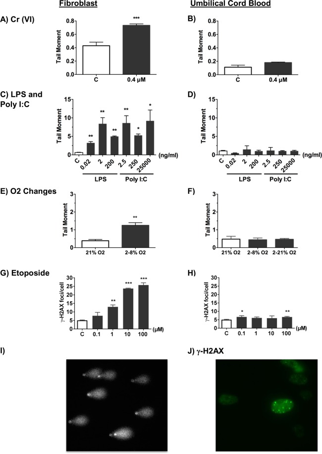
Figure 1.
Differential DNA damage response between fibroblasts and cord blood after exposure to conditioned media from BeWo barriers. The level of DNA damage in fibroblasts as recorded by either the mean tail moment, using the alkaline comet assay (A–F), or immunocytochemical analysis of the mean number of γ-H2AX foci per cell (DNA double-strand breaks). (G,H) Values are shown for fibroblasts (left hand column) and umbilical cord blood (UCB) mononuclear cells (right hand column) after a 24 hour exposure to conditioned media below BeWo barriers resting on transwell inserts. The top surface of BeWo barriers was exposed to culture media without (open histograms) or with (shaded histograms) (A,B) 0.4 uM Cr(VI); (C,D) LPS or PolyI:C (ng/ml); (E,F) altered levels of oxygen; (G,H) etoposide (0.1, 1, 10 and 100 μM). Representative images of (I) comet assay and (J) γ-H2AX immunostaining are shown. Plots represent the mean ± SD of 3 independent experiments. n = 3. *p < 0.05, **p < 0.01, ***p < 0.001 as determined by unpaired two-tailed student’s t-test.

