Fig. 5.
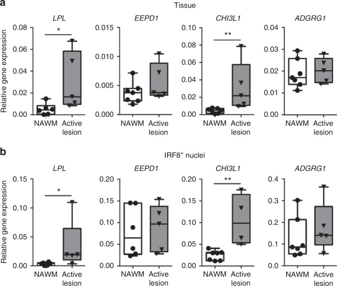
Gene expression differences observed in normal-appearing white matter (NAWM) microglia relate to changes found in multiple sclerosis (MS) lesions. a Excised mixed active/inactive MS lesion tissue (n = 5) shows increased expression of the differentially expressed genes LPL and CHI3L1 compared to NAWM (n = 7) determined by quantitative reverse transcription polymerase chain reaction (RT-qPCR). EEPD1 and ADGRG1 expression is unaltered. Mann–Whitney U test: *p < 0.05, **p < 0.01. b IRF8+ nuclei isolated from the same mixed active/inactive MS lesions demonstrates increased expression of CHI3L1 and LPL, compared to IRF8+ nuclei from NAWM, determined by RT-qPCR. In line with the tissue data, the expression of EEPD1 and ADGRG1 is not changed in IRF8+ nuclei from MS lesions. Mann–Whitney U one-tailed test: *p < 0.05, **p < 0.01. Box plot center line shows median; whiskers show minimum–maximum values

