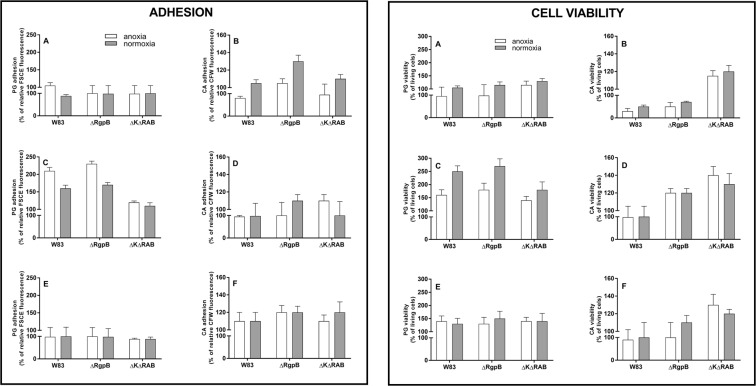
Figure 1.
Adhesion (left panel) and viability (right panel) of P. gingivalis (PG) and C. albicans (CA) cells that form a dual-species biofilm (A,B) Sequential model type I. A biofilm was formed by CFSE-stained bacteria for 24 h at 37 °C in Schaedler medium under anoxic conditions and this mono culture was covered with C. albicans cells and further propagated for 3 h at 37 °C in RPMI medium under both anoxic and normoxic conditions. After biofilm washing, the fungal cells were visualized by CFW staining. Adhesion was determined fluorometrically. Cell viability was determined after the biofilm had formed by cell scratching and propagation on the agar plate in media and under conditions appropriate for each species. (C,D) Sequential model type II. A biofilm was formed by C. albicans cells for 24 h at 37 °C in RPMI medium under normoxic conditions, and then settled with bacteria for 3 h at 37 °C in RPMI medium under anoxic and normoxic conditions. Further analyses were performed as described in (A,B). (E,F) Simultaneous model. A dual-species biofilm was formed by bacterial and fungal contact at the cell surface at the same time for 3 h at 37 °C in RPMI medium under anoxic and normoxic conditions. Further analyses were performed as described in A and B. All experiments (A–F) were performed with five repetitions in triplicate. The data shown are relative to a mono-species biofilm propagated under the same conditions. Data were presented as means ± SD (standard deviation) and analyzed using GraphPad Prism software (GraphPad, LaJolla, CA).

