Figure 4.
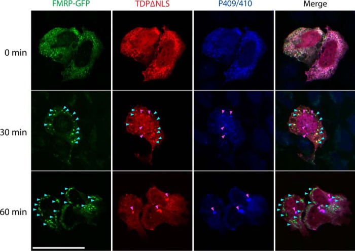
TDP-43 aggregates undergo a dynamic transition from cytoplasmic granules to mature inclusions. Cells co-expressing cytoplasmic TDP-43 (TDP-43–ΔNLS) and FMRP–GFP were treated with 0.25 mm arsenite for up to 60 min, followed by fixation and triple labeling to detect FMRP granules (green), total TDP-43 (red), and pathological phospho-TDP-43–positive (P409/410) inclusions (blue) by confocal microscopy. Cyan arrowheads highlight FMRP granules, and magenta arrowheads highlight mature TDP-43 inclusions. We note a gradual stress-dependent transition in TDP-43 morphology from granular SG-associated puncta to mature inclusions by ∼30 min, which corresponded with the emergence of phosphorylated TDP-43 (P409/410) inclusions and minimal co-localization with FMRP–GFP. Scale bar, 20 μm.

