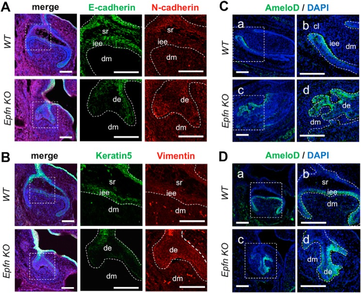
Figure 7.
Epfn deficiency induces the partial epithelial-mesenchymal transition in the invading epithelia. A, immunofluorescence of E-cadherin and N-cadherin in E16 WT and Epfn-KO molars. Green, E-cadherin; red, N-cadherin; blue, DAPI. B, immunofluorescence of keratin 5 and vimentin in E16 WT and Epfn-KO molars. Green, keratin 5; red, vimentin; blue, DAPI. C, immunofluorescence of AmeloD in E17 WT and Epfn-KO incisors. b and d, enlargements of a and c. D, immunofluorescence of AmeloD in E17 WT and Epfn-KO molars. b and d, enlargements of a and c. Green, AmeloD; blue, DAPI. de, dental epithelium; dm, dental mesenchyme; iee, inner enamel epithelium; sr, stellate reticulum. Scale bars, 100 μm.

