Figure 8.
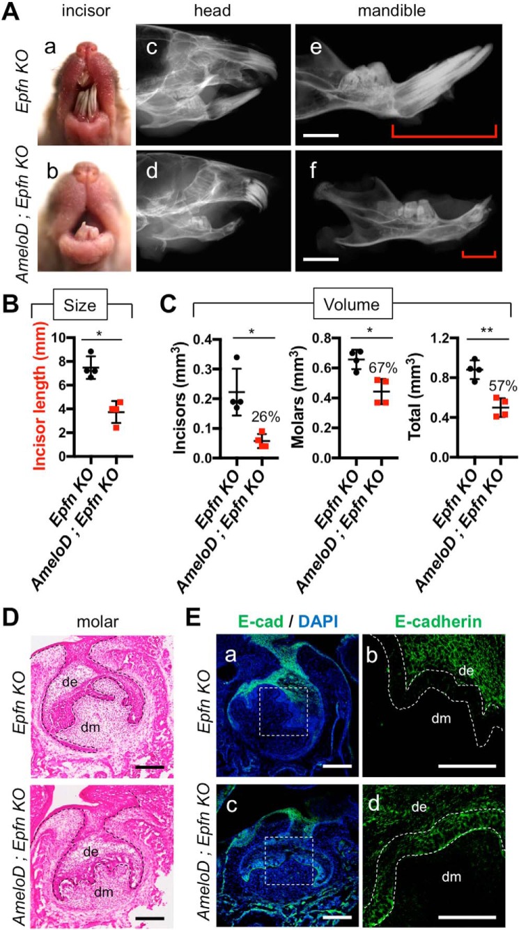
AmeloD deficiency reduces the tooth size and number in Epfn-KO mice via inhibition of random dental epithelial invasions. A, photographic analyses (a and b) and radiographic analysis (c–f) of 3-month-old Epfn-KO and AmeloD; Epfn-KO heads. The red brackets indicate the length of the incisors. Scale bars, 1000 μm. B, incisor lengths of 3-month-old Epfn-KO and AmeloD; Epfn-KO mice (n = 5). The mean is shown as lines. Error bars represent S.D. *, p < 0.05 with a two-tailed t test. C, the total volume of dentin in 6-week-old Epfn-KO and AmeloD; Epfn-KO teeth by micro-CT analysis. Left panel, total volume of incisors. Middle panel, total volume of molars. Right panel, total volume of molars and total volume of incisor were quantified. The ratio of the volume (AmeloD; Epfn KO/Epfn KO) is shown as the number above the bar graph (n = 4). The mean is shown as lines. Error bars represent S.D. *, p < 0.05; **, p < 0.01 with a two-tailed t test. D, H-E staining of P1 Epfn-KO and AmeloD; Epfn-KO molars. The dashed lines indicate the border between the dental epithelium and mesenchyme. Scale bars, 100 μm. E, immunofluorescence staining analysis of E-cadherin (E-cad) in P1 Epfn-KO and AmeloD; Epfn-KO molars. b and d, enlargements of a and c. Green, E-cadherin; blue, DAPI. The dashed lines indicate the presumptive inner enamel epithelium. de, dental epithelium; dm, dental mesenchyme. Scale bars, 100 μm.

